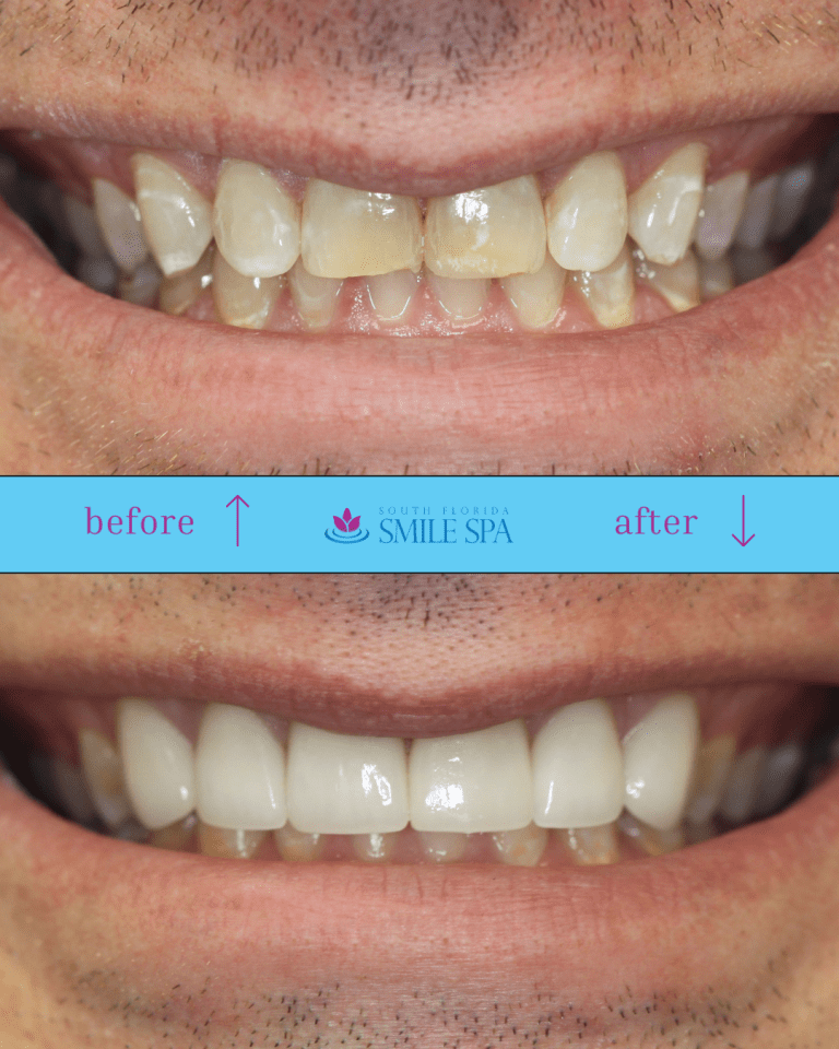


Dental Implant Safety in Pompano Beach: What a 3D Scan Sees That an X-Ray Cannot
Quick answer: A cone beam CT (CBCT) scan is a low-dose, three-dimensional X-ray of your jaw. It measures the height, width and density of the bone where an implant will go, and it shows the exact position of the nerve canal and the floor of your sinus — none of which a flat X-ray can measure.
The American Academy of Oral and Maxillofacial Radiology recommends cross-sectional imaging for the assessment of every dental implant site and names CBCT the imaging method of choice. At South Florida Smile Spa in Pompano Beach, Dr. Nicole M. Berger, DDS scans before every implant case. The reason is blunt: in the published data, implants placed within one millimeter of the nerve canal caused lasting numbness in roughly two out of three patients, while implants kept two millimeters away caused none.
This guide covers dental implant safety Pompano Beach patients should expect: bone, nerve, sinus, radiation dose, and the questions worth asking before surgery.

By Dr. Nicole M. Berger, DDS · South Florida Smile Spa · Pompano Beach, FL
The question a flat X-ray cannot answer
Patients ask me a version of the same question every week. Why do I need another scan? You already took X-rays.
It is a fair question, and the answer is not “because the technology is newer.” It is because a flat X-ray and a 3D scan answer different questions.
A traditional dental X-ray — a periapical or a panoramic — is a shadow. Everything between the X-ray source and the sensor is flattened into a single image, front to back, with no depth. That is genuinely useful for what it was designed to do: find decay between teeth, check the length of a root, look at bone levels, spot an abscess.
But an implant is not a two-dimensional decision. Placing one means drilling a channel into living bone that has a nerve running through it, a sinus cavity sitting above it, and neighboring tooth roots on either side. The three questions that decide whether that is safe are all depth questions:
- How thick is the bone from cheek side to tongue side? A flat film cannot show width at all.
- How far is the crest of the bone from the nerve canal? A panoramic image can suggest this, and it can be wrong by millimeters because of magnification and angle.
- Is the bone actually solid, or is it a thin shell around a hollow? Density does not read reliably off a shadow.
A flat X-ray can tell me a site looks plausible. It cannot tell me a site is safe. Those are not the same standard, and for surgery the second one is the only one that counts.
What cone beam 3D imaging actually is
Cone beam computed tomography is an X-ray technique that captures your anatomy volumetrically instead of as a flat projection. The scanner arm rotates once around your head — you sit or stand still, mouth closed, nothing enters your mouth, nothing touches you — and takes a rapid series of images from many angles. Software reconstructs those into a 3D volume of your jaws.
From that volume I can do things a flat film will never allow:
- Slice through your jaw at any point and look at a true cross-section, cheek-to-tongue
- Measure in millimeters in any direction, without magnification distortion
- Trace the path of the inferior alveolar nerve through the mandible and see exactly where it runs beneath the site
- Rotate the jaw and look at the implant site from the angle the drill will actually approach it
- Plan the implant virtually — length, width, angle, depth — before a single instrument is opened
The scan itself takes seconds. The planning that follows takes considerably longer, and that is where its value lives.
The cone beam imaging technology in our Pompano Beach office is part of a broader digital workflow, alongside intraoral scanning and same-day restorative technology. You can see the full list on our technology page.
Dental implant safety Pompano Beach: what I look for when I read your scan
A scan is not a photograph you glance at. Reading one is a systematic process, and here is what I am actually working through on an implant case:
The nerve. In the lower jaw, the inferior alveolar nerve travels through a bony canal. It supplies sensation to your lip, chin, and lower teeth. I trace its full course and establish exactly how much vertical bone sits above it at the planned site.
The sinus. In the upper back jaw, the maxillary sinus sits directly above the molar roots. When an upper molar has been missing for a while, the sinus floor often drops. I measure how much bone remains beneath it, because that number determines whether the site needs augmentation.
Bone width. Bone resorbs from the outside in after a tooth is lost. A ridge can look tall and adequate on a panoramic film and be a knife-edge in cross-section — too narrow to hold an implant without grafting.
Bone density. Different regions of the jaw have very different bone quality. Density influences implant selection, drilling protocol, and whether the implant can be loaded immediately or needs time to integrate.
Lingual concavity. The inner surface of the lower jaw is often scooped inward. A drill angled as if the jaw were a straight block can perforate the tongue-side plate. This is invisible on any flat image and obvious in cross-section.
Adjacent roots. Implants need clearance from neighboring teeth. Roots curve in ways that flat films disguise.
Anything that should not be there. Retained root fragments, cysts, chronic infection, unexpected anatomy.
Seven questions. One scan answers all of them before anyone picks up an instrument.
The millimeter that decides everything
If you take one number away from this article, take this one.
A systematic review and meta-analysis published in Medicina Oral Patología Oral y Cirugía Bucal grouped implant cases by how close the implant ended up to the mandibular canal, then looked at how many patients developed neurosensory problems — numbness, tingling, or altered sensation in the lip and chin.
| Distance from implant to nerve canal | Reported neurosensory alteration |
|---|---|
| 2 mm or more | 0% |
| 1–2 mm | 0% |
| Less than 1 mm | 68% |
| Implant intruding into the canal | 53% |
Read that table again. The difference between a completely uneventful outcome and a two-in-three chance of a lasting sensory change is roughly the thickness of a credit card and a half.
That is the entire argument for 3D imaging, stated in one line. Nobody plans to place an implant into a nerve. It happens when the planning was done on an image that could not resolve the difference between one millimeter and two. Surgeons commonly work to a safety margin of about 2 mm above the canal — and you cannot honor a 2 mm margin using a measurement tool with more error than that.
The same logic applies upward into the sinus, sideways into the lingual plate, and laterally into the roots of neighboring teeth. Implant complications are overwhelmingly planning failures, not hand-skill failures.
Is the radiation safe? Here are the actual numbers
This deserves a straight answer rather than reassurance, because a cone beam scan does deliver more radiation than a standard dental X-ray. Anyone who tells you otherwise is glossing.
Radiation dose is measured in microsieverts (µSv). Direct comparison studies of dental imaging put the typical figures roughly here:
| Imaging type | Typical effective dose | Roughly equal to |
|---|---|---|
| Single intraoral X-ray | ~1.5 µSv | A few hours of ordinary living |
| Panoramic X-ray | ~22 µSv | About 2–3 days |
| Dental cone beam CT | ~61–134 µSv | About 1–2 weeks |
| Medical CT of the jaws | Several hundred to over 1,000 µSv | Weeks to months |
The “roughly equal to” column is background radiation — the dose every one of us absorbs simply from existing on this planet, from soil, building materials, food and cosmic rays. It runs around 2,400 µSv per year, which works out to roughly 8.5 µSv per day.
So: a dental cone beam scan is meaningfully more than a single X-ray, dramatically less than a medical CT of the same region, and in absolute terms lands somewhere near one to two weeks of the radiation you were going to absorb anyway.
The honest framing is not “it is nothing.” The honest framing is that this is a dose worth accepting when it changes the surgical plan, and not worth accepting when it does not. Which brings us to the part most technology articles skip.
The Lead Apron Question — and Why We Still Use One
You may have read that dental offices are doing away with the lead apron. Nationally, that is true, and it is worth explaining properly — including why you will still be handed one in our office.
In 2024 the ADA published its recommendations on radiation safety and regulatory issues in dental imaging and concluded that thyroid collars and lead abdominal aprons are no longer necessary, building on patient shielding recommendations the American Academy of Oral and Maxillofacial Radiology issued in 2023. Two things drove that conclusion. Modern digital equipment delivers a small fraction of the dose the machines those aprons were designed for delivered. And the beam is now collimated tightly to the area being imaged, so a correctly positioned apron is not intercepting a great deal.
There is a second consideration that gets less attention. A shield placed carelessly can drift into the diagnostic beam and obscure part of the image. When that happens the image has to be retaken, which means a second exposure. That is a legitimate concern, and it is as much an argument for positioning a shield properly as it is for removing it.
Here is where I land. “No longer necessary” is not the same thing as “no longer permitted.” We continue to provide a lead apron at South Florida Smile Spa, and we have no plans to change that. Some patients simply want one. Some are anxious about imaging and find the weight of it steadying. Placed correctly, it does not interfere with the image at all. I would rather a patient sit through a scan feeling covered than technically optimized and uneasy.
What I do want you to understand is where your protection actually comes from: equipment that runs at a fraction of the dose it used to, a beam narrowed to the site being examined, and — the largest factor by far — only taking the image when it is going to change what I do. The apron was never doing most of that work. It was only the most visible part of it.
What the ADA Changed in January 2026
For a long time, the authoritative guidance on when to take a 3D scan came from the AAOMR rather than from the ADA itself. That changed this year.
On 5 January 2026 the American Dental Association published its first dental X-ray patient-selection recommendations in more than a decade — and the first from the ADA ever to address 3D cone beam imaging alongside conventional 2D. They were developed by an expert panel convened by the ADA Council on Scientific Affairs, endorsed by the AAOMR, and published in the January issue of the Journal of the American Dental Association, with an ahead-of-print version in Oral Surgery, Oral Medicine, Oral Pathology and Oral Radiology.
The headline principle is the one I have practiced under for years: order imaging only when it is clinically necessary, after reviewing the patient’s medical and dental history, any prior images, and the findings of an actual examination. The panel’s lead author put it in terms I would use myself — you would not have any other part of your body imaged unless a clinician had examined you first and had a reason.
What makes this document useful rather than merely reassuring is that it works through specific clinical scenarios rather than issuing a general principle and stopping. Implant planning and placement is one of them. So are cracked teeth, endodontic cases, and temporomandibular disorders.
Read alongside the AAOMR position statement described in the next section, the two are consistent rather than in tension. The ADA’s broader rule is that cone beam imaging belongs where lower-exposure options cannot supply the diagnostic information required. For an implant site, they cannot — bone width and nerve position are cross-sectional facts, and a flat image is structurally incapable of measuring them. The guidance asks me to justify the scan. For an implant, the justification is the first half of this article.
When a 3D scan is the right call — and when it is not
The professional guidance here is specific, and I follow it.
The American Academy of Oral and Maxillofacial Radiology, in its position statement on imaging for dental implants, recommends that the radiographic examination of any potential implant site include cross-sectional imaging, and that CBCT be considered the imaging method of choice for obtaining it. It also flags situations where 3D imaging is particularly indicated — sinus augmentation, bone grafting, assessment of impacted teeth in the field of interest, and evaluation of prior traumatic injury.
Just as importantly, the same body sets limits:
- Not as a first look. For the initial evaluation of a patient considering implants, panoramic imaging supplemented by intraoral periapicals is the appropriate starting point. Cross-sectional imaging is not recommended as an initial examination.
- Not for routine implant check-ups. In the absence of symptoms, periapical radiographs are what is indicated for postoperative monitoring of implants. CBCT is not for periodic review of implants that are doing fine.
- Not without the training to read it. The guidance is explicit that clinicians must be competent to interpret the images they take — including normal anatomy, anatomic variants, and signs of disease outside the immediate area of interest.
A practice that scans everyone who walks through the door, for everything, is not being thorough. It is being indiscriminate, and radiation is not free. The standard I hold to is the one the evidence supports: image when the image will change what I do.
What else the scan sees: your mouth is not a closed system
Here is the part of cone beam imaging that patients almost never anticipate, and that I think is genuinely the most important thing on this page.
A 3D scan of your jaws does not stop politely at your teeth. Depending on the field of view, it captures the sinuses, the airway, part of the cervical spine, and the soft tissues of the neck — including the region where the carotid artery divides.
In a study of 400 large-field CBCT scans published in the International Journal of Dentistry, researchers found 653 incidental findings in 309 of the 400 scans — an average of about two per scan. Airway findings appeared in 11.49% of scans. Calcification in the external carotid artery appeared in 10.41%. And roughly 31% of the findings identified were judged to require referral to another clinician.
Calcification in the carotid artery is not a dental finding. It is a marker of atherosclerosis — the same disease process that produces heart attacks and strokes. When it turns up on a scan taken to plan an implant, the correct response is a phone call to the patient’s physician, not a note in the dental chart.
This is the same thread that runs through everything I write here. The American Dental Association’s MouthHealthy resource is direct about the fact that oral health is connected to conditions well beyond the mouth, including cardiovascular disease and diabetes. We cover that link in our guide to deep cleaning and gum disease, and it applies here in an unusually literal way — the imaging we take to solve a dental problem sometimes sees the beginning of a medical one.
To be clear about what this is and is not: a dental cone beam scan is not a cardiac screening test, and it cannot diagnose carotid disease. Doppler ultrasound does that, ordered by a physician. What a scan can do is notice something and start a conversation that would not otherwise have happened. That is not a small thing.
From scan to surgical guide
The scan is data. The plan is what you do with it.
Once I have the 3D volume, I place your implant virtually — selecting size, position, depth and angle in software, checking clearance to the nerve, the sinus, the lingual plate and the adjacent roots from every direction. Only when the position is right on screen does anything happen in the operatory.
That plan can then be transferred into a surgical guide: a physical template that seats over your teeth or ridge and constrains the drill to the position that was planned. The virtual plan and the actual surgery become the same event.
The practical consequences for you are shorter surgical time, smaller incisions in many cases, and a restoration designed around the implant position from the outset rather than compromised to fit wherever the implant ended up. It also means the conversation we have beforehand is concrete. I can show you your own anatomy on screen and explain exactly why the plan is what it is — which is a very different experience from being told to trust the process.
If you are still weighing whether an implant is the right answer at all, our comparison of dental implants versus a bridge works through that decision, and a smile makeover consultation covers the broader planning process when more than one tooth is involved.
A composite patient scenario
Composite scenario. Assembled from common clinical patterns to illustrate a point. This is not a real patient, and it is not a testimonial or a representation of any individual’s results.
Consider a patient in her late fifties who lost a lower right first molar about three years ago and has finally decided to replace it. Her panoramic X-ray looks straightforward. There is apparently ample bone height above the nerve canal, and nothing about the film suggests any complication.
The cone beam scan tells a longer story. Vertical bone above the canal measures adequately — but the ridge has resorbed to a narrow crest, too thin buccolingually for a standard-diameter implant without augmentation. There is also a pronounced lingual concavity below the crest, meaning a drill angled the way the flat film would have suggested would have headed toward the tongue-side plate rather than through solid bone. And in the sinus above, an unrelated mucous retention cyst — harmless, but worth documenting.
None of that was visible on the panoramic image. None of it made the case impossible. What it did was change the plan: a graft to rebuild width, a different implant diameter, a corrected angle of approach, and a conversation about timeline that happened before treatment started rather than in the middle of it.
That is what the scan buys. Not drama. Just the absence of surprises.
If the machine itself is what worries you
For some patients, the imaging is the anxious part — the equipment, the stillness, the sense of being processed by something.
The scan is brief, open, and nothing goes in your mouth. You are not enclosed the way a medical CT or MRI encloses you. Most people find it the easiest part of the visit.
But if dental anxiety is what has kept you from addressing a missing tooth for years, the scan is not really the obstacle — the whole appointment is. That is worth naming, because it is solvable. Dr. Berger personally administers and monitors IV sedation, with continuous vital-sign monitoring throughout, and a great many of our implant patients complete their treatment under sedation dentistry rather than white-knuckling through it. Our guide to the six real fears anxious patients bring to a sedation dentist addresses this directly.
Imaging precision and sedation are the same argument from two directions. Both exist so that a difficult procedure becomes an uneventful one.
Five questions to ask before implant surgery anywhere
Whether you are considering treatment with us or with any other practice, these are worth asking:
- Will you take a cone beam 3D scan before placing my implant? If the answer is no, ask how bone width and nerve position will be determined without one.
- Who reads the scan, and what training do they have in interpreting it? Taking an image and reading it competently are separate skills.
- Will you show me my own scan and walk me through the plan? You are entitled to see the reasoning, not just the conclusion.
- How much clearance will there be between the implant and the nerve? A practice planning in 3D can answer this in millimeters.
- What happens if the scan shows something unexpected? The answer should include a referral pathway, not just a dental one.
A practice that welcomes those questions is telling you something useful. So is one that deflects them. Our article on choosing a dentist in Pompano Beach expands this into a fuller checklist.
Frequently asked questions
Does a cone beam scan hurt?
No. Nothing enters your mouth and nothing touches you. You rest your chin on a small support, hold still, and the scanner arm rotates around your head. The scan portion typically takes well under a minute, and the exposure itself is a matter of seconds.
How long does a 3D dental scan take?
The scan is a few seconds of exposure within an appointment slot of a few minutes, including positioning. The images are available immediately — there is no waiting period and no separate imaging center to visit. The planning work that follows is what takes real time, and it happens after you have gone home.
Is cone beam imaging safe during pregnancy?
Elective dental imaging is generally deferred during pregnancy, and elective implant surgery usually is too. Tell us if you are pregnant or think you might be, and we will discuss timing. Urgent situations are assessed individually with appropriate shielding and the smallest field of view that answers the clinical question — but “we can wait” is very often the right answer.
Can I use a cone beam scan from another dentist, or a CT my physician already ordered?
Sometimes. A recent dental CBCT from another practice can often be used if the field of view covers the site and the image quality is adequate — bring the data file, not printouts. A medical CT is a different matter: it may cover the anatomy, but medical scans are captured at different resolutions and settings and are frequently not suitable for implant planning. We would rather review what you have than repeat imaging unnecessarily.
Do I need a 3D scan for a single implant, or only for full-arch cases like All-on-4?
Both. The professional recommendation is cross-sectional imaging for the assessment of every implant site, not only complex ones. A single lower molar sits closer to the inferior alveolar nerve than almost any other implant position — it is not the simple case people assume it is.
What happens if the scan finds something unrelated to my teeth?
We tell you, we document it, and where appropriate we refer you to your physician or the relevant specialist. Most incidental findings turn out to be benign and need nothing more than a note. A minority — sinus disease, airway narrowing, arterial calcification — warrant a medical opinion. Either way, you find out.
Is cone beam imaging used for anything besides dental implants?
Yes. It is valuable in evaluating complex root canal anatomy, assessing impacted teeth before extraction, investigating jaw joint problems, measuring bone loss from advanced periodontal disease, and diagnosing pain that flat imaging cannot explain. It is a diagnostic tool, not an implant-only tool — but the same rule applies throughout: it is warranted when the image will change the decision.
Do you still use a lead apron for dental X-rays?
Yes. We provide a lead apron at South Florida Smile Spa and have no plans to stop. National guidance did change: the ADA’s 2024 radiation safety recommendations, building on shielding recommendations from the American Academy of Oral and Maxillofacial Radiology in 2023, concluded that thyroid collars and lead abdominal aprons are no longer necessary, because modern digital equipment uses a small fraction of the dose older machines delivered and the beam is collimated tightly to the area being imaged. But “no longer necessary” is not the same as “not permitted,” and many patients find the apron reassuring. Positioned correctly it does not interfere with the image, so we keep it.
The bottom line
Implant dentistry has an unusual property: the difference between an excellent outcome and a serious complication is often established before the surgery begins, in the planning. A cone beam scan is how that planning stops being an estimate.
Two millimeters of clearance from the nerve, versus one. That is what is being decided while you are sitting still for fifteen seconds with a scanner arm moving around your head.
If you are considering an implant in Pompano Beach and want a plan built on your actual anatomy rather than an educated guess, we would be glad to talk it through.
South Florida Smile Spa · Nicole M. Berger, DDS
572 E McNab Rd, Suite 102, Pompano Beach, FL 33060
(954) 785-1100
Hours: Monday 8:30 AM–12:30 PM · Tuesday 8:00 AM–5:00 PM · Wednesday 8:30 AM–12:30 PM · Thursday 9:30 AM–6:30 PM · Friday 8:00 AM–5:00 PM · Saturday and Sunday closed
This article is for general educational purposes and does not constitute individual dental or medical advice. Imaging decisions, treatment recommendations and outcomes vary by patient and require an in-person examination.
Plan your implant on your actual anatomy.
Cone beam 3D imaging and guided planning with Dr. Nicole M. Berger, DDS in Pompano Beach.
See Our 3D Imaging TechnologyRequest a Consultation
Or call (954) 785-1100
Sources
- American Academy of Oral and Maxillofacial Radiology — Position statement on selection criteria for the use of radiology in dental implantology, with emphasis on cone beam computed tomography
- Inferior alveolar nerve damage related to dental implant placement: a systematic review and meta-analysis, Medicina Oral Patología Oral y Cirugía Bucal
- Patient radiation dose and protection from cone-beam computed tomography, Imaging Science in Dentistry
- Barghan S, Tetradis S, Mallya SM et al. — Incidental findings on cone beam computed tomography studies outside of the maxillofacial skeleton, International Journal of Dentistry
- American Dental Association — MouthHealthy

Dr. Nicole M. Berger, DDS
Dr. Berger has cared for patients in Pompano Beach for over two decades at South Florida Smile Spa, with a focus on cosmetic dentistry, implants and reconstruction, and sedation options for anxious patients.